Migraine is a major global health problem that affects over 10% of the population.1,2
Although it's a distinct disease, a wide range of factors and pathological mechanisms can
contribute to the development of migraine in an individual, which can be affected by, for
example, genetic, environmental, and metabolic factors.3 The occurrence of a migraine
attack can be also influenced by factors such as elevation in specific neuropeptide
levels.3 The appearance of specific symptoms and clinical features during an attack,
for example whether a patient experiences aura or nausea or photophobia, may depend on genetic,
anatomical, as well as neurophysiological and other factors.3 Over time, and with
advances in neuroimaging techniques, it's now understood that migraine is a complex neurological
disease that is associated with changes in biology, as well as in the structure and function in
the central nervous system.3,4
So, there have been new insights into migraine pathophysiology that have contributed to our
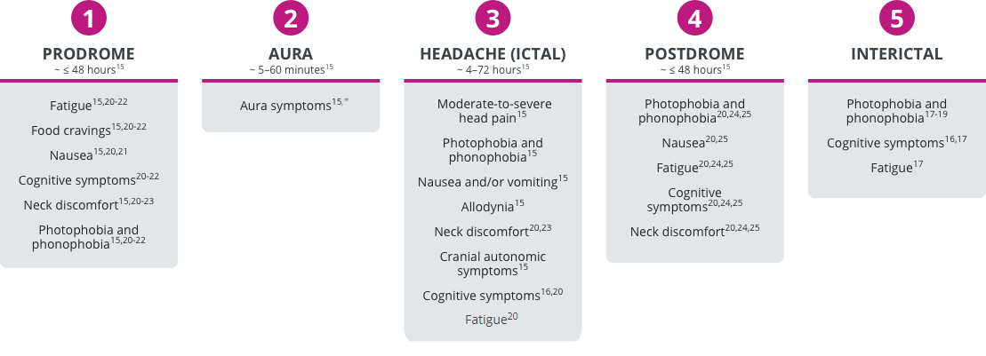
evolving understanding of this disease.4,5 Migraine attacks occur over hours to days
and are divided into several phases characteristically.6-8 Those include the
prodrome, the aura, headache, and the postdrome, as well as the interictal state, where patients
may still continue to experience symptoms of migraine even in the absence of
headache6-8. Some of those non-pain symptoms particularly during the prodromal phase
can include fatigue, cognitive symptoms, photophobia, phonophobia, osmophobia, and other sensory
sensitivities.6,9,10 Nonpain symptoms associated with a headache phase
characteristically and classically include photophobia, phonophobia, nausea, and
vomiting.6 And, attacks, of course, can be associated with symptoms of cutaneous
allodynia, usually in the form of scalp sensitivity that clinically manifests by patients where
they will say they don't like a cap on their head or a ponytail or glasses sitting on the bridge
of their nose.6,11 That's characteristic of hypersensitivity to sensory stimulation
in the skin, otherwise known as allodynia.11 Functional imaging studies have
demonstrated that pain and non-pain symptoms experienced during the various phases of migraine
may be associated with specific brain regions.5,7 For example, the hypothalamus is
associated with some of the common prodromal symptoms, such as fatigue, photophobia,
phonophobia, and cognitive symptoms that appear during a prodrome and have been shown to be
present in the earliest phases of an attack, when the first anatomical site to be activated is
indeed the hypothalamus.5,12,13
The potential clinical and pathophysiological consequences of uncontrolled migraine include
increased frequency of headache or migraine days over time, changes in the type of symptoms that
are experienced by patients, changes in the severity of symptoms, as well as changes in the
consumption of medication, and, indeed, in the effectiveness of medications.13-16 As
an example of disease progression, patients can transition from episodic to chronic migraine
over time.13,17 The incidence of that progression is about 3% per year.13
Now, there are a number of factors that have been associated with that progression from episodic
to higher frequency migraine, then ultimately to chronic migraine.18 The most
important of which is the baseline headache frequency, as well as the frequency with which
individuals use acute medications.18 But, there are other factors that have been
shown independently to increase the risk of progression, including the presence of cutaneous
allodynia, the presence of nausea, and, indeed, the effectiveness of acute
medication.18 So, the less optimal the responses to an acute medicine, the more
likely that individual is to progress over time, highlighting the importance of optimizing the
acute therapy of individual attacks.19 Migraine frequency can also improve over time
with a higher proportion of patients transitioning from chronic migraine to episodic migraine
than from episodic to chronic migraine.17,20 So, that's the good news that there is a
significant proportion of patients who do remit to episodic migraine over time.20
And, sometimes, those patients will move back and forth from an episodic to a chronic pattern,
indicating the dynamic nature of this disease.21
Now, there are potential clinical and pathophysiological consequences of uncontrolled migraine,
as well, that include the overuse of acute medication, and we all know that patients who overuse
medication, usually defined as more than ten days per month, can develop what's known as
medication overuse headache, which is a common problem occurring in about 1-2% of the
population.6,22 Imaging data suggests that patients on the more severe end of the
disease spectrum are associated with more pronounced neurological sequalae, and there have been
structural and functional alterations seen on functional neuroimaging in patients with longer
disease duration, in patients who have a higher headache frequency, and in patients with
medication overuse.23-28 For example, structural imaging studies in patients with
migraine suggests that there is a correlation between increased headache-day frequency and
alterations in grey matter volume.23
Now, the clinical assessment of migraine severity can be achieved in a variety of different
ways.6,29 We can evaluate the headache-day frequency and the consumption of acute
medications with the use of a diary, for example6. We can use patient-reported
outcome tools to assess the functional impairment of patients over time.29 We can
track non-pain symptoms over time, both during the attacks and in between, or in the interictal
phase.30-34 And, of course, taking a comprehensive history of patients, including
comorbidities.35
References:
- GBD 2017 Disease and Injury Incidence and Prevalence Collaborators. Lancet. 2018;
392:1789–1858.
- Stovner LJ, et al. Cephalalgia. 2007; 27:193–210.
- Charles A. Lancet Neurol. 2018; 17(2):174-182.
- Tfelt-Hansen PC, et al. J Headache Pain. 2011; 51:752-778.
- Schwedt TJ, Chong CD. Curr Opin Neurol. 2015; 28(3):265–270.
- Headache Classification Committee of the International Headache Society (IHS) The
International Classification of Headache Disorders, 3rd edition. Cephalalgia. 2018;
38(1):1–211.
- Goadsby PJ, et al. Physiol Rev. 2017; 97:553–622.
- Houtveen JH, Sorbi MJ. PLoS One. 2013; 8(8):e72827.
- Giffin NJ, et al. Neurology. 2003; 60:935–940.
- Schulte LH, et al. J Headache Pain. 2015; 16:14.
- Burstein R, et al. Ann Neurol. 2000; 47:614-624.
- Maniyar FH, et al. Brain. 2014; 137; 232–241.
- Bigal ME, Lipton RB. Curr Neurol Neurosci Rep. 2011; 11:139–148.
- Bigal ME, et al. Neurology. 2008; 70(17):1525–1533.
- Negro A, et al. Curr Treat Options Neurol. 2017; 19:32.
- Diener HC, Limmroth V. Lancet Neurol. 2004; 3:475–483.
- Bigal ME, et al. Headache. 2008; 48:1157-1168.
- Buse DC, et al. Headache. 2019; 59:306-338.
- Lipton RB, et al. Headache. 2016; 56:1635-1648.
- Manack A, et al. Neurology. 2011; 76:711–718.
- Serrano D, et al. J Headache Pain. 2017; 18:101.
- Kristoffersen ES, Lundqvist C. J Pain Res. 2014; 7:367–378.
- Kim JH, et al. Cephalalgia. 2008; 28:598–604.
- Schwedt TJ, et al. Cephalalgia. 2014; 34(12):947–958.
- Chong CD, Schwedt TJ. Cephalalgia. 2015; 35(13):1162-1171.
- Zhao L, et al. J Headache Pain. 2013; 38(1):1–211.
- Chen Z, et al. J Headache Pain. 2017; 18:112.
- Fumal A, et al. Brain. 2006; 129:543–550.
- Buse DC, et al. Mayo Clin Proc. 2009; 84(5):422-435.
- Lipton RB, et al. Headache. 2012; 53:81-92.
- Nasreddine ZS, et al. J Am Geriatr Soc. 2005; 53:695–699.
- Buysee DJ, et al. Psychiatry Res,. 1988; 28:193-213.
- Rosti-Otajarvi E, et al. Brain Behav. 2017; 7:e00743.
- Lipton RB, et al. Ann Neurol. 2003; 63(2):148–158.
- Weatherall MW. Ther Adv Chronic Dis. 2015; 6(3):115–123